From conception to birth to growing through those ups and downs of infancy, toddlerhood, childhood and the infamous teenage… a child is what makes a parent’s life worth every struggle.
Imagine however, the despondence, the despair, the helplessness, the anger, the confusion when the angel arrives carrying, along with other blessings, defects in body.
Some manifest at birth, some gradually as the child comes of age. We will delve upon various facets of Congenital Anomalies in this article, being carried in three parts.
Congenital anomalies in humans present in a myriad of ways. Many of these are manageable, even treatable, early detection and intervention being the game changer. It’s as surprising as it is saddening to see the extent of lack of awareness in our society, not only in the section of society where one would expect it but in affluent educated classes and even the medical fraternity.
In over a decade of working in paediatric rehabilitation, I’ve come across incidences where even after a year at rehabilitation, the parents don’t even know what the underlying processes of their child’s problems are. In this direction it’s a small endeavour to provide some insight into the basics of major congenital and developmental anomalies.
Congenital means acquired in the womb. Congenital anomalies can be defined as structural or functional anomalies that occur during intrauterine life and are present at birth. Also called birth defects, congenital disorders, or congenital malformations, these conditions develop prenatally and may be identified before or at birth, or later in life.
GLOBAL INCIDENCE
About 30 to 70/1000 live births. In India – 2.5 to 4 % of children are born with abnormalities Most common type of birth defect being-CNS abnormalities (22%) resulting in hundreds of thousands of associated deaths.
However, the true number of cases may be much higher because statistics do not often consider terminated pregnancies and stillbirths.
Congenital anomalies are one of the main causes of the global burden of disease, and low- and middle-income countries are disproportionately affected. These areas are also less likely to have facilities to treat reversible conditions, leading to more pronounced and long-lasting effects.
TYPES
Birth defects may result in disabilities that may be physical, intellectual, or developmental. The disabilities can range from mild to severe.
Birth defects are divided into two main types: structural disorders in which problems are seen with the shape of a body part and functional disorders in which problems exist with how a body part works, how a person learns, or the senses.
Functional disorders include metabolic and degenerative disorders.Some congenital anomalies have both structural and developmental effects. Examples of these include fragile X syndrome, spina bifida, and Down syndrome.
STRUCTURAL ANOMALIES
Some common structural congenital anomalies include heart defects, spina bifida, a cleft lip or palate, and clubfoot.
Heart defects
The most common congenital anomalies are heart defects. Most have no obvious cause, but if a pregnant woman has diabetes or smokes during pregnancy, it may increase the chance.
A heart anomaly occurs when part of the heart does not form properly in the womb. This can affect how well it can circulate blood around the body.There are many different types of heart defects; depending on the affected area of the heart.
The most common heart defect is a ventricular septal defect. This is a hole in the wall separating the two lower heart chambers. Sometimes, the hole will close on its own over time. Infants with severe heart defects often need surgery soon after birth.
Limb reduction
Sometimes, part of a limb will not form completely in the womb. Known as limb reduction, this structural anomaly means that a limb is smaller than the usual size or missing altogether. For example, an infant may have a missing finger, clubfoot, or an arm that is shorter than usual.
People with a minor limb reduction may find that it does not affect daily life. However, others may find certain movements limited, more difficult, or not possible. Physical therapy, splints, or prosthetics can help.
The cause of limb reduction is unknown. Exposure to chemicals or infections during pregnancy might increase the chance.
Cleft lip or palate
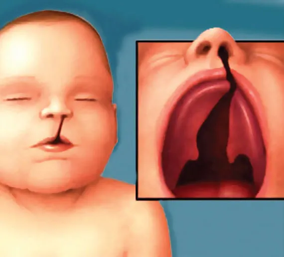
If the tissues forming the roof of the mouth or lip do not join properly, it can cause a cleft lip or palate. Some infants may have both.
This can affect speech, hearing, and eating. Most infants with this structural anomaly will need surgery in the first few months of life. In severe cases, they may need ongoing surgeries.
Neural tube defects
Neural tube defects affect the brain and spinal cord. These structural anomalies occur in the first few months of pregnancy, when the brain and spinal cord of a fetus are forming.
Types of neural tube defects include:
Anencephaly: This occurs when parts of the brain and skull do not form at all.
Encephalocele: This occurs when the neural tube does not close fully. The brain projects through an opening in the skull.
Spina bifida: This occurs when the spine does not form and close properly. This affects the nerves and spinal cord.
Both anencephaly and encephalocele are very rare. Spina bifida is more common, affecting 1-2 cases in every 1000 live births globally and 4-8 every 100 live births in India.
The symptoms of spina bifida can be mild or severe, depending on the part of the spine it affects. It can cause paralysis, learning difficulties, and bladder and bowel problems.
Taking folic acid during pregnancy can help prevent neural tube defects in the infant.
Gut and stomach anomalies
Sometimes, the stomach muscles do not form properly and leave a hole near the belly button. This can mean that the intestines or organs are outside of the body.
In gastroschisis, the abdominal wall does not completely close, so the bowel can push through and develop outside of the body. The organs will not have a protective sac. In omphalocele, which is associated with other anomalies, there is a protective sac around the organs.
In either case, an infant will need surgery soon after birth.
The muscle separating the chest and abdomen is called the diaphragm. If a hole forms in the diaphragm, the organs can begin to move into the chest. This is known as a diaphragmatic hernia.
An infant will need surgery and help to breathe until the lungs work normally.
The author is Physiotherapist, In-charge PMRD, Khyber Medical Institute
Disclaimer: The views and opinions expressed in this article are the personal opinions of the author.
The facts, analysis, assumptions and perspective appearing in the article do not reflect the views of GK.


